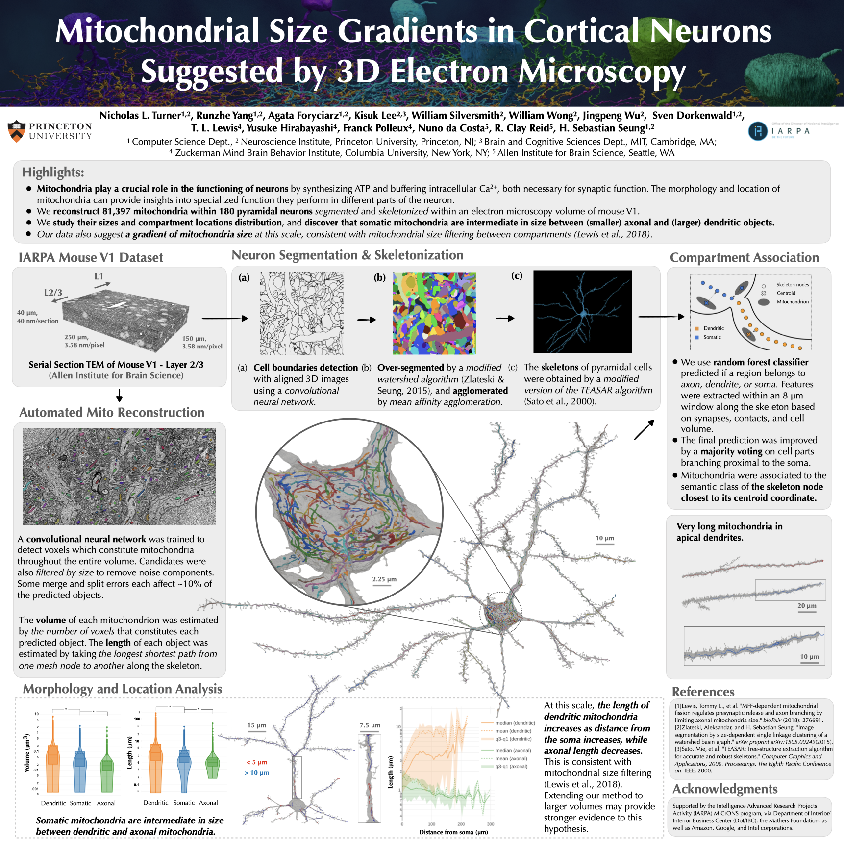
Mitochondria play a crucial role in the functioning of neurons by synthesizing ATP and buffering intracellular Ca2+, both necessary for synaptic function. The morphology and location of mitochondria can provide insights into specialized function they perform in different parts of the neuron. We reconstruct 81,397 mitochondria within 180 pyramidal neurons segmented and skeletonized within an electron microscopy volume of mouse V1. We study their sizes and compartment locations distribution, and discover that somatic mitochondria are intermediate in size between (smaller) axonal and (larger) dendritic objects. Our data also suggest a gradient of mitochondria size at this scale, consistent with mitochondrial size filtering between compartments (Lewis et al., 2018).
Abstract
Neurons are among the most highly polarized cells in our body and this includes compartmentalized organelle structure and function. Mitochondria play a crucial role in the functioning of neurons by synthesizing ATP and buffering intracellular Ca2+, both necessary for synaptic function. They are highly dynamic organelles, are constantly changing through fusion and fission events to adapt to changing environment in the cell (Youle et al. 2012), moving across cells (Narayanareddy et al. 2014) and often forming long filaments (Popov et al. 2005, Amchenkova et al. 1988). The morphology and location of mitochondria can provide insights into specialized function they perform in different parts of the neuron. Observations of neuronal mitochondria thus far have mainly been in dissociated neurons in vitro, single neurons in vivo with light microscopy, or small volumes (< 1000 um3) with 3D electron microscopy (EM). Recently Dorkenwald et al. (2017) demonstrated automated reconstruction of mitochondria from 3D EM of millions of um3 of avian brain. Such large-scale reconstructions open up the possibility of obtaining more detailed and quantitative information about either cell-type specific and/or compartment-specific mitochondrial morphology and distribution. We acquired 3D EM images of mouse primary visual cortex (V1). All neurons with somas in the imaged volume were reconstructed using a semiautomated pipeline described elsewhere. We applied a 3D convolutional network to classify voxels as either mitochondrion or background. The mitochondrial voxels were grouped into over 900,000 mitochondria using connected components. For subsequent analyses, we focused on the mitochondria that were contained in neurons with somas in the volume. Each mitochondrion was assigned to either axon, dendrite, or soma. We calculated the distance from soma centroid to each mitochondrion centroid. It is well-known that axonal mitochondria are typically shorter and smaller than dendritic mitochondria. Our data show that somatic mitochondria are intermediate in size between axonal and dendritic mitochondria. This comparison may be simplistic, as mitochondria vary in size systematically with distance from the soma. We found that the size (length and volume) of axonal mitochondria decreases with distance from the soma. In contrast, the size of dendritic mitochondria increases with distance from the soma up to almost 100 microns. Our data so far strongly suggest that the balance between mitochondrial fission and fusion must be compartment-specific with fusion dominating over fission in dendrites and vice versa in axons.
Poster @ Princeton AIML Retreat

Authors
Nicholas L. Turner, Runzhe Yang, Agata Foryciarz, Kisuk Lee, William Silversmith, William Wong, Jingpeng Wu, Sven Dorkenwald, T. L. Lewis, Yusuke Hirabayashi, Franck Polleux, Nuno da Costa, R. Clay Reid, H. Sebastian Seung